Research
Questions
I study how plants use calcium signaling to sense and respond to their environment, and how stress can disrupt these signals. I approach plant immunity through the lens of calcium signaling, using advanced biosensor imaging, molecular biology, biochemistry, and genetics to uncover how immune responses are regulated.


Whole-plant calcium imaging highlights signaling
diversity based on immune receptor activated during ETI.
Calcium signaling and bacterial growth at two temperatures. At elevated temperature (dotted line), the RPS4-dependent calcium signal fails to reach the immunity threshold, leading to ETI collapse and increased pathogen growth.
How Does Elevated Temperature Suppress Calcium Signaling During ETI?
Whole-plant calcium imaging shows that elevated temperature dampens the calcium influx normally triggered during effector-triggered immunity (ETI). This suppression correlates with a loss of resistance, but the underlying mechanisms are just now being resolved. Does temperature directly affect NLR receptor activity, and can we find mutations to confer temperature resilience for climate reilience immunity?
How is Immunity-Related Calcium Signaling Controlled Within Cells and Organelles?
Using advanced biosensor imaging, I track calcium dynamics at cellular and subcellular resolution during effector-triggered immunity (ETI). Several open question remain including 1) where does NLR-mediated calcium influx initiate within the cell, and 2) how are calcium signaling and immune outputs are coordinated between cells at the site of effector recognition and those in surrounding tissues?

Subcellular calcium signaling dynamics in an epidermal pavement cell during the onset of ETI

Temporal color coding tracks peak calcium levels
to illustrate spatiotemporal calcium signaling heterogeneity in the epidermis during ETI.

Ratiometric calcium imaging experiments show
baseline calcium levels are suppressed
after incubation at elevated temperature.

The virulent pathogen DC3000 actively suppresses host cellular calcium in an effector-dependent manner compared to the effectorless strain Δ36E.
How Do Elevated Temperature and Pathogens Alter Calcium Homeostasis?
Elevated temperature suppresses steady-state calcium, while pathogen effectors actively reduce host calcium during infection. What mechanisms do pathogens use to suppress calcium availability, and how does temperature rewire the calcium homeostatic system?
How does calcium misregulation shift plant–microbe relationships?
Arabidopsis aca4/11 mutants lack two vacuolar calcium pumps, leading to spontaneous calcium waves and temperature-dependent lesions. As seedlings, these plants show heightened resistance to pathogens, yet as they mature they permit commensal microbes to overgrow, resulting in a dysbiotic community. This dual phenotype links disrupted calcium clearance not only to cell death and lesioning but also to the balance between protective immunity and microbial homeostasis.
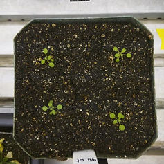

23 °C
28 °C
aca4/11 mutants (left) and wildtype (right) grown at two different temperatures. aca4/11 plants exhibit spontaneous lesions, dwarfism, and autoimmunity at 23 °C.
Rosette leaf of an aca4/11 plant grown at 28 °C, then shifted to 23 °C immediately before imaging. Calcium wavefronts (segmented in green) can be seen. Areas with persistently elevated calcium precede lesion formation.


Plant-wide calcium signaling in response to a shift to high humidity.

Kymographic analysis highlights spatiotemporal patterns of calcium waves in the petioles in response to high humidity.
How Does Calcium Signaling Control Molecular and Physiological Responses to Humidity Change?
Climate-controlled imaging reveals that shifts in humidity trigger distinct calcium signatures, from rapid bursts to sustained waves. These signals are required for adaptive leaf movement, yet their mechanistic basis is still being resolved. How do cell wall integrity sensors and calcium channels integrate to mediate plant responses to fluctuating humidity?
How does microbial signaling and metabolism change during an infection?
Using genetically-encoded flourescent protein-based biosensors, we have the capability to track changes spatiotemporal changes in bacterial pathogen signaling and metabolism in real time at the population and individual levels. These experiments will allow us to glean insights into crucial events expereienced by the pathogen that can steer an infection towards success or failure in myriad plant species and under different environmental conditions.

Pseudomonas syringae (Pst) was infiltrated into tobacco leaf tissue grown under different environmental conditions. Depending on the condition, biosensor signal from the bacteria changes in flourescent intensity, indicating differential physiological changes in the bacterial pathogen during infection. chlA (chlorophyll A) is in magenta and Pst biosensor bacteria are in green.
In Preparation
Publications
2023
2022
2021
2020
2018
2017
2016
2014